1. Introduction
The measurement and detection of ionizing radiation constitute a fundamental component of nuclear science and engineering. Radiation detection systems are designed to identify the presence of radiation, quantify its intensity, and, in many cases, determine its energy distribution. Because different types of radiation possess distinct physical properties, their interactions with matter and the methods required for their detection differ significantly.
Ionizing radiation commonly encountered in nuclear engineering includes alpha particles (α), beta particles (β), gamma rays (γ), X-rays, and neutrons. These radiations differ in several important aspects, including:
- mass and electric charge
- energy range
- interaction mechanisms with matter
- penetration capability
- spectral characteristics
Understanding these differences is essential for selecting appropriate radiation detectors and for interpreting radiation measurements in applications such as nuclear power systems, radiation protection, environmental monitoring, nuclear security, and nuclear medicine.
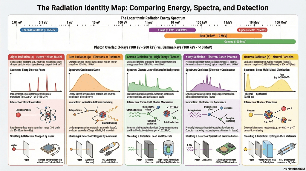
Figure 1 (Radiation Identity Map) illustrates the relationship between these radiation types by comparing their characteristic energy ranges, spectral signatures observed in detectors, and typical detection technologies. The upper part of the diagram presents a logarithmic radiation energy scale spanning from approximately 0.01 eV to 100 MeV, which demonstrates that ionizing radiation covers more than ten orders of magnitude in energy. Within this range, different radiation types occupy distinct regions, although some overlap occurs, particularly between X-rays and gamma rays.
- 2. Classification of Ionizing Radiation
Ionizing radiation can be broadly categorized into two groups based on whether the radiation carries electric charge.
Charged Particle Radiation
Charged particle radiation includes alpha particles and beta particles, which carry electric charge and interact strongly with matter through Coulomb forces. As these particles pass through matter, they lose energy continuously through ionization and excitation of atoms. Because of these strong interactions, charged particles deposit energy over relatively short distances.
Charged particles therefore produce direct ionization tracks, which makes them relatively easy to detect using detectors that measure ionization or scintillation produced along the particle trajectory.
Neutral Radiation
Neutral radiation includes gamma rays, X-rays, and neutrons, which do not carry electric charge. These radiations interact with matter through different physical mechanisms and generally exhibit much greater penetration than charged particles.
Gamma rays and X-rays are forms of electromagnetic radiation, consisting of high-energy photons. Neutrons, in contrast, are massive neutral particles produced in nuclear reactions such as fission or fusion.
Because neutral radiation does not interact through direct Coulomb forces, it cannot produce ionization directly. Instead, these radiations must first generate secondary charged particles before they can be detected. For photons, these secondary particles are typically electrons produced by photon interactions, while neutrons must undergo nuclear reactions that emit charged particles.
- 3. Alpha Radiation
Alpha particles consist of helium nuclei containing two protons and two neutrons. They are typically emitted during the radioactive decay of heavy nuclei such as uranium, radium, plutonium, and americium. Because of their relatively large mass and positive charge, alpha particles interact strongly with matter and lose energy rapidly.
Energy Spectrum
Alpha particles are usually emitted with discrete energies, corresponding to specific nuclear transitions. As a result, alpha radiation spectra typically exhibit sharp monoenergetic peaks, which are clearly illustrated in the radiation identity map.
Typical alpha particle energies range from approximately 4 to 9 MeV, depending on the radionuclide. For example:
- Americium-241 emits alpha particles with an energy of approximately 5.49 MeV
- Polonium-210 emits alpha particles with energies near 5.30 MeV
Because the alpha particle energy distribution is narrow, alpha spectroscopy using semiconductor detectors can achieve excellent energy resolution, allowing precise identification of alpha-emitting isotopes.
Range and Penetration
Despite their relatively high energies, alpha particles have very limited penetration due to their strong interactions with matter. Their typical ranges are:
- a few centimeters in air
- approximately 20–80 micrometers in solid materials
Consequently, alpha radiation can be easily stopped by a thin sheet of paper or by the outer layer of human skin.
Detection Methods
Alpha radiation is commonly measured using detectors that respond to the intense ionization produced by the particle. Typical alpha detectors include:
- silicon semiconductor detectors
- gas proportional counters
- zinc sulfide scintillation detectors
Because alpha particles have such short ranges, these detectors often require the radioactive source to be positioned very close to the detector surface or measured in vacuum chambers.
- 4. Beta Radiation
Beta radiation consists of high-energy electrons or positrons emitted during nuclear beta decay. In contrast to alpha decay, beta decay involves the transformation of a neutron into a proton or vice versa, accompanied by the emission of a beta particle and a neutrino.
Energy Spectrum
A defining characteristic of beta radiation is its continuous energy spectrum, which is illustrated in the infographic as a broad distribution curve. The kinetic energy released during beta decay is shared between the emitted beta particle and the neutrino. As a result, beta particles can be emitted with any energy between zero and a characteristic maximum endpoint energy.
Typical beta endpoint energies range from tens of keV to several MeV. Examples include:
- Carbon-14: endpoint energy of 156 keV
- Strontium-90: endpoint energy of 546 keV
- Phosphorus-32: endpoint energy of 1.71 MeV
The continuous nature of beta spectra provides an important diagnostic feature for identifying beta-emitting radionuclides.
Range and Penetration
Beta particles penetrate matter more deeply than alpha particles but less deeply than gamma rays. Their ranges typically extend:
- several meters in air
- a few millimeters in tissue or solids
When beta particles interact with high-atomic-number materials, they may produce bremsstrahlung radiation, which consists of secondary X-rays generated when the charged particle decelerates in the electric field of atomic nuclei.
Detection Methods
Common detectors used for beta radiation include:
- Geiger–Müller counters
- plastic scintillation detectors
- semiconductor detectors
Because beta spectra are continuous, specialized techniques such as beta spectrometry may be used to analyze their energy distributions.
- 5. Gamma Rays and X-Rays
Gamma rays and X-rays are both forms of electromagnetic radiation, differing primarily in their origin. Gamma rays originate from nuclear transitions, whereas X-rays are typically produced through atomic electron transitions or bremsstrahlung processes.
Energy Range
Gamma rays generally possess higher energies than most X-rays. Typical ranges include:
- X-rays: approximately 1 keV – 200 keV
- Gamma rays: approximately 100 keV – several MeV
Examples of common gamma-ray energies include:
- Cesium-137: 662 keV
- Cobalt-60: 1.17 MeV and 1.33 MeV
The infographic also illustrates an important concept: X-ray and gamma energy ranges overlap, meaning that the distinction between them is based primarily on their origin rather than energy alone.
Interaction with Matter
Gamma rays and X-rays interact with matter primarily through three mechanisms:
- Photoelectric absorption
- Compton scattering
- Pair production
The dominant interaction mechanism depends strongly on photon energy and the atomic number of the absorbing material.
Gamma-Ray Energy Spectrum
Gamma-ray spectroscopy typically reveals characteristic spectral features such as:
- photopeaks
- Compton continua
- Compton edges
- backscatter peaks
These features arise from the various photon interaction processes occurring within the detector material.
For example:
- photopeaks occur when the photon deposits its entire energy in the detector
- Compton continua arise from partial energy transfer during Compton scattering
- pair production becomes possible at photon energies above 1.022 MeV
Detection Methods
Photon radiation is commonly detected using:
- scintillation detectors such as NaI(Tl) or CsI
- semiconductor detectors such as HPGe (High-Purity Germanium)
- cadmium telluride or silicon detectors for X-ray detection
Semiconductor detectors are particularly valuable for spectroscopy because of their superior energy resolution.
- 6. Neutron Radiation
Neutrons are electrically neutral particles that interact with matter primarily through nuclear reactions rather than electromagnetic interactions. Because they lack electric charge, neutrons do not directly ionize atoms and must therefore be detected indirectly.
Energy Spectrum
Neutron energies span an extremely wide range, commonly classified into several categories:
| Neutron Type | Energy |
| Thermal neutrons | ~0.025 eV |
| Epithermal neutrons | 0.5 eV – 10 keV |
| Fast neutrons | 10 keV – 10 MeV |
| High-energy neutrons | >10 MeV |
In nuclear reactors, the neutron energy spectrum typically contains both a thermal peak and a fast neutron component, resulting in a broad distribution such as that illustrated in the infographic.
Detection Mechanisms
Neutron detection relies on nuclear reactions that produce charged particles. Common reactions include:


The charged particles generated in these reactions are then detected through conventional ionization processes.
Neutron Detectors
Common neutron detectors include:
- helium-3 proportional counters
- boron trifluoride detectors
- lithium-glass scintillators
- organic scintillators for fast neutrons
Because neutrons are highly penetrating, their detection often requires moderation materials such as polyethylene or water to slow fast neutrons to thermal energies.
- 7. Comparison of Radiation Energy Ranges
The approximate energy ranges of the major radiation types can be summarized as follows:
| Radiation Type | Typical Energy Range |
| X-rays | 1 keV – 200 keV |
| Gamma rays | 100 keV – several MeV |
| Beta particles | tens of keV – several MeV |
| Alpha particles | 4 – 9 MeV |
| Neutrons | 0.025 eV – tens of MeV |
These large differences in energy and interaction mechanisms explain why different radiation types require different detection techniques and shielding strategies.
- 8. Summary
Different types of ionizing radiation exhibit distinct physical properties that influence both their interaction with matter and the methods used for their detection.
Charged particles such as alpha and beta radiation produce ionization directly and can therefore be detected through relatively straightforward ionization measurements. In contrast, photons such as gamma rays and X-rays must first produce secondary electrons within the detector material before they can be measured. Neutrons require an additional step, involving nuclear reactions that generate detectable charged particles.
A fundamental principle in radiation detection can therefore be summarized as follows:
Charged particles are detected directly through ionization, photons are detected indirectly through secondary electrons, and neutrons are detected through nuclear reactions.
Understanding these distinctions is essential for the design and operation of radiation detection systems used in nuclear engineering, radiation protection, environmental monitoring, nuclear security, and medical applications.
- References
- Knoll, G. F. (2010). Radiation Detection and Measurement (4th ed.). John Wiley & Sons.
- Turner, J. E. (2007). Atoms, Radiation, and Radiation Protection (3rd ed.). Wiley.
- Gilmore, G. (2008). Practical Gamma-Ray Spectrometry (2nd ed.). Wiley.
- L’Annunziata, M. F. (2012). Handbook of Radioactivity Analysis (3rd ed.). Academic Press.
- Leo, W. R. (1994). Techniques for Nuclear and Particle Physics Experiments. Springer.
###############################################################
- ประเภทของรังสี สเปกตรัมพลังงาน และหลักการตรวจวัด
- 1. บทนำ
การตรวจวัดและการวัดปริมาณรังสีไอออไนซ์เป็นองค์ประกอบพื้นฐานที่สำคัญของวิทยาศาสตร์และวิศวกรรมนิวเคลียร์ ระบบตรวจวัดรังสีมีหน้าที่ตรวจสอบการมีอยู่ของรังสี วัดความเข้มของรังสี และในหลายกรณีสามารถวิเคราะห์การกระจายตัวของพลังงานของรังสีได้ด้วย เนื่องจากรังสีแต่ละชนิดมีสมบัติทางกายภาพแตกต่างกัน กลไกการเกิดอันตรกิริยากับสสารและวิธีการตรวจวัดจึงแตกต่างกันอย่างมีนัยสำคัญ
รังสีไอออไนซ์ที่พบได้บ่อยในงานวิศวกรรมนิวเคลียร์ประกอบด้วย
- อนุภาคแอลฟา (α)
- อนุภาคบีตา (β)
- รังสีแกมมา (γ)
- รังสีเอกซ์ (X-ray)
- นิวตรอน
รังสีแต่ละชนิดแตกต่างกันในหลายด้าน เช่น
- มวลและประจุไฟฟ้า
- ช่วงพลังงาน
- กลไกการเกิดอันตรกิริยากับสสาร
- ความสามารถในการทะลุผ่านวัสดุ
- ลักษณะของสเปกตรัมพลังงาน
การเข้าใจความแตกต่างเหล่านี้มีความสำคัญต่อการเลือกใช้ หัววัดรังสี ที่เหมาะสม รวมถึงการตีความผลการตรวจวัดรังสีในงานด้านต่าง ๆ เช่น โรงไฟฟ้านิวเคลียร์ การป้องกันอันตรายจากรังสี การติดตามกัมมันตภาพรังสีในสิ่งแวดล้อม ความมั่นคงปลอดภัยทางนิวเคลียร์ และเวชศาสตร์นิวเคลียร์
ภาพประกอบ “แผนภาพอัตลักษณ์ของรังสี” แสดงความสัมพันธ์ระหว่างชนิดของรังสี ช่วงพลังงาน ลักษณะสเปกตรัม และวิธีการตรวจวัด โดยแกนด้านบนของภาพเป็น สเกลพลังงานแบบลอการิทึม ครอบคลุมตั้งแต่ประมาณ 0.01 อิเล็กตรอนโวลต์ (eV) จนถึง 100 เมกะอิเล็กตรอนโวลต์ (MeV) ซึ่งแสดงให้เห็นว่ารังสีไอออไนซ์ครอบคลุมช่วงพลังงานกว้างมากกว่า สิบลำดับขนาด
- 2. การจำแนกประเภทของรังสีไอออไนซ์
โดยทั่วไปสามารถจำแนกรังสีไอออไนซ์ออกเป็นสองกลุ่มหลักตามการมีประจุไฟฟ้า
รังสีชนิดมีประจุ
รังสีชนิดมีประจุประกอบด้วย อนุภาคแอลฟาและอนุภาคบีตา อนุภาคเหล่านี้มีประจุไฟฟ้าและเกิดอันตรกิริยากับสสารผ่านแรงไฟฟ้าระหว่างอนุภาคมีประจุกับอิเล็กตรอนในอะตอม เมื่ออนุภาคเคลื่อนที่ผ่านสสารจะสูญเสียพลังงานอย่างต่อเนื่องผ่านกระบวนการ
- การแตกตัวเป็นไอออน
- การกระตุ้นอะตอม
เนื่องจากอันตรกิริยาเกิดขึ้นอย่างรุนแรง อนุภาคมีประจุจึงสูญเสียพลังงานอย่างรวดเร็วและมีระยะทางเคลื่อนที่สั้นในสสาร
รังสีชนิดไม่มีประจุ
รังสีชนิดไม่มีประจุประกอบด้วย
- รังสีแกมมา
- รังสีเอกซ์
- นิวตรอน
รังสีแกมมาและรังสีเอกซ์เป็น รังสีแม่เหล็กไฟฟ้า ส่วน นิวตรอน เป็นอนุภาคที่ไม่มีประจุไฟฟ้าแต่มีมวล เนื่องจากไม่มีประจุไฟฟ้า รังสีเหล่านี้ไม่สามารถก่อให้เกิดการแตกตัวเป็นไอออนโดยตรงได้ แต่ต้องเกิดกระบวนการที่ทำให้เกิด อนุภาคมีประจุทุติยภูมิ ก่อนจึงจะตรวจวัดได้
ตัวอย่างเช่น
- โฟตอนของรังสีแกมมาหรือรังสีเอกซ์จะทำให้เกิดอิเล็กตรอนพลังงานสูงในตัวตรวจวัด
- นิวตรอนต้องเกิด ปฏิกิริยานิวเคลียร์ ที่สร้างอนุภาคมีประจุ
- 3. รังสีแอลฟา
อนุภาคแอลฟาเป็น นิวเคลียสของฮีเลียม ซึ่งประกอบด้วยโปรตอนสองตัวและนิวตรอนสองตัว อนุภาคชนิดนี้มักเกิดจากการสลายตัวของธาตุกัมมันตรังสีที่มีเลขอะตอมสูง เช่น ยูเรเนียม เรเดียม หรือพลูโทเนียม เนื่องจากมีมวลมากและมีประจุบวกสองหน่วย อนุภาคแอลฟาจึงเกิดอันตรกิริยากับสสารอย่างรุนแรงและสูญเสียพลังงานอย่างรวดเร็ว
สเปกตรัมพลังงาน
อนุภาคแอลฟามักถูกปล่อยออกมาด้วย ค่าพลังงานเฉพาะเจาะจง ซึ่งสัมพันธ์กับระดับพลังงานของนิวเคลียส ดังนั้นสเปกตรัมของรังสีแอลฟาจึงปรากฏเป็น ยอดพลังงานแหลมที่มีค่าคงที่
ช่วงพลังงานของอนุภาคแอลฟาโดยทั่วไปอยู่ระหว่างประมาณ4 – 9 MeV
ตัวอย่างเช่น
- อะเมริเซียม-241 ปล่อยอนุภาคแอลฟาที่ประมาณ 5.49 MeV
- โพโลเนียม-210 มีพลังงานประมาณ 5.30 MeV
เนื่องจากสเปกตรัมมีความแคบ การวิเคราะห์สเปกตรัมของรังสีแอลฟาจึงสามารถทำได้ด้วยความละเอียดสูงโดยใช้ หัววัดรังสีชนิดสารกึ่งตัวนำ
ระยะทางและการทะลุผ่าน
แม้ว่าจะมีพลังงานสูง แต่อนุภาคแอลฟามีความสามารถในการทะลุผ่านต่ำมาก เนื่องจากเกิดอันตรกิริยากับสสารอย่างรุนแรง
ระยะทางโดยทั่วไปคือ
- ประมาณ 3–8 เซนติเมตรในอากาศ
- ประมาณ 20–80 ไมโครเมตรในของแข็ง
ดังนั้นแผ่นกระดาษบาง ๆ หรือแม้แต่ผิวหนังชั้นนอกก็สามารถหยุดอนุภาคแอลฟาได้
วิธีการตรวจวัด
หัววัดที่ใช้ตรวจวัดรังสีแอลฟา ได้แก่
- หัววัดกึ่งตัวนำซิลิคอน
- หัววัดก๊าซแบบสัดส่วน
- หัววัดซินทิลเลเตอร์สังกะสีซัลไฟด์
เนื่องจากอนุภาคแอลฟามีระยะทางสั้น แหล่งกำเนิดรังสีจึงต้องอยู่ใกล้กับหัววัดมาก หรือวัดในสภาวะสุญญากาศ
- 4. รังสีบีตา
รังสีบีตาประกอบด้วย อิเล็กตรอนหรือโพซิตรอนพลังงานสูง ที่เกิดจากการสลายตัวของนิวเคลียส ในกระบวนการนี้นิวตรอนสามารถเปลี่ยนเป็นโปรตอน หรือโปรตอนเปลี่ยนเป็นนิวตรอน พร้อมกับการปล่อยอนุภาคบีตาและนิวตริโน
สเปกตรัมพลังงาน
ลักษณะเด่นของรังสีบีตาคือ สเปกตรัมพลังงานต่อเนื่อง
พลังงานที่เกิดจากการสลายตัวถูกแบ่งระหว่าง
- อนุภาคบีตา
- นิวตริโน
ดังนั้นพลังงานของอนุภาคบีตาจึงมีค่าได้ตั้งแต่ศูนย์จนถึงค่าพลังงานสูงสุดที่เรียกว่า พลังงานปลายสเปกตรัม
ตัวอย่างเช่น
- คาร์บอน-14 : 156 keV
- สตรอนเชียม-90 : 546 keV
- ฟอสฟอรัส-32 : 1.71 MeV
ระยะทางและการทะลุผ่าน
อนุภาคบีตาสามารถทะลุผ่านสสารได้มากกว่าแอลฟา แต่ยังน้อยกว่ารังสีแกมมา
ระยะทางโดยทั่วไป
- หลายเมตรในอากาศ
- หลายมิลลิเมตรในเนื้อเยื่อ
เมื่ออนุภาคบีตาผ่านวัสดุที่มีเลขอะตอมสูง อาจเกิด รังสีเบรมสตราห์ลุง (Bremsstrahlung) ซึ่งเป็นรังสีเอกซ์ทุติยภูมิที่เกิดจากการชะลอตัวของอนุภาคมีประจุ
วิธีการตรวจวัด
หัววัดที่ใช้ตรวจวัดรังสีบีตา ได้แก่
- หัววัดไกเกอร์–มึลเลอร์
- หัววัดซินทิลเลเตอร์พลาสติก
- หัววัดกึ่งตัวนำ
- 5. รังสีแกมมาและรังสีเอกซ์
รังสีแกมมาและรังสีเอกซ์เป็นรังสีแม่เหล็กไฟฟ้าที่มีพลังงานสูง ความแตกต่างหลักอยู่ที่ แหล่งกำเนิด
- รังสีแกมมาเกิดจากการเปลี่ยนระดับพลังงานของนิวเคลียส
- รังสีเอกซ์เกิดจากกระบวนการของอิเล็กตรอนในอะตอม
ช่วงพลังงาน
ช่วงพลังงานโดยทั่วไปคือ
รังสีเอกซ์ ประมาณ 1 keV – 200 keV
รังสีแกมมา ประมาณ 100 keV – หลาย MeV
ตัวอย่างพลังงานรังสีแกมมา
- ซีเซียม-137 : 662 keV
- โคบอลต์-60 : 1.17 และ 1.33 MeV
ควรสังเกตว่า ช่วงพลังงานของรังสีเอกซ์และรังสีแกมมาซ้อนทับกันได้ ดังนั้นการจำแนกจึงอาศัย แหล่งกำเนิดของรังสีเป็นหลัก
อันตรกิริยากับสสาร
โฟตอนพลังงานสูงเกิดอันตรกิริยากับสสารผ่านสามกลไกหลัก
- ปรากฏการณ์โฟโตอิเล็กทริก
- การกระเจิงแบบคอมป์ตัน
- การเกิดคู่อนุภาค
- การชนแล้วทำให้นิวเคลียสแตกตัว (กรณีโฟตอนพลังงานสูงมาก ๆ หลักสิบ MeV)
กลไกที่เกิดขึ้นเด่นชัดขึ้นอยู่กับ
- พลังงานของโฟตอน
- เลขอะตอมของวัสดุ
สเปกตรัมของรังสีแกมมา
สเปกตรัมของรังสีแกมมาที่วัดด้วยหัววัดมักประกอบด้วยองค์ประกอบสำคัญหลายส่วน ได้แก่
- ยอดพลังงานเต็ม
- พื้นหลังคอมป์ตัน
- ขอบคอมป์ตัน
- ยอดกระเจิงย้อนกลับ
องค์ประกอบเหล่านี้สะท้อนกระบวนการอันตรกิริยาของโฟตอนภายในตัวตรวจวัด
วิธีการตรวจวัด
หัววัดที่ใช้ตรวจวัดโฟตอน ได้แก่
- หัววัดซินทิลเลเตอร์ เช่น โซเดียมไอโอไดด์
- หัววัดกึ่งตัวนำเจอร์เมเนียมบริสุทธิ์สูง
- หัววัดกึ่งตัวนำแคดเมียมเทลลูไรด์หรือซิลิคอนสำหรับรังสีเอกซ์
หัววัดกึ่งตัวนำมีข้อได้เปรียบคือ ความละเอียดพลังงานสูง
- 6. รังสีนิวตรอน
นิวตรอนเป็นอนุภาคไม่มีประจุที่เกิดอันตรกิริยากับสสารผ่าน ปฏิกิริยานิวเคลียร์ แทนที่จะเป็นอันตรกิริยาไฟฟ้าแม่เหล็ก
ช่วงพลังงาน
พลังงานของนิวตรอนครอบคลุมช่วงกว้างมาก
| ประเภทนิวตรอน | พลังงาน |
| นิวตรอนความร้อน | ~0.025 eV |
| นิวตรอนกึ่งความร้อน | 0.5 eV – 10 keV |
| นิวตรอนเร็ว | 10 keV – 10 MeV |
| นิวตรอนพลังงานสูง | >10 MeV |
ในเครื่องปฏิกรณ์นิวเคลียร์ สเปกตรัมของนิวตรอนมักมีทั้ง ยอดพลังงานความร้อนและส่วนของนิวตรอนเร็ว
กลไกการตรวจวัด
การตรวจวัดนิวตรอนอาศัยปฏิกิริยานิวเคลียร์ที่สร้างอนุภาคมีประจุ เช่น

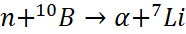

อนุภาคมีประจุที่เกิดขึ้นจะถูกตรวจวัดผ่านกระบวนการไอออไนเซชัน
หัววัดนิวตรอน
ตัวอย่างหัววัดนิวตรอน ได้แก่
- หัววัดก๊าซฮีเลียม-3
- หัววัดโบรอนไตรฟลูออไรด์
- หัววัดซินทิลเลเตอร์ลิเทียม
- หัววัดซินทิลเลเตอร์อินทรีย์สำหรับนิวตรอนเร็ว
เนื่องจากนิวตรอนทะลุผ่านได้ดี จึงมักใช้วัสดุที่มีไฮโดรเจนสูง เช่น พอลิเอทิลีนหรือน้ำ เพื่อชะลอนิวตรอนก่อนตรวจวัด
- 7. การเปรียบเทียบช่วงพลังงานของรังสี
| ชนิดของรังสี | ช่วงพลังงานโดยประมาณ |
| รังสีเอกซ์ | 1 keV – 200 keV |
| รังสีแกมมา | 100 keV – หลาย MeV |
| อนุภาคบีตา | หลาย keV – หลาย MeV |
| อนุภาคแอลฟา | 4 – 9 MeV |
| นิวตรอน | 0.025 eV – หลายสิบ MeV |
ความแตกต่างของช่วงพลังงานและกลไกอันตรกิริยาเป็นเหตุผลสำคัญที่ทำให้ต้องใช้ วิธีการตรวจวัดและวัสดุป้องกันที่แตกต่างกัน
- 8. สรุป
รังสีไอออไนซ์แต่ละชนิดมีสมบัติทางกายภาพที่แตกต่างกัน ซึ่งส่งผลต่อกลไกการเกิดอันตรกิริยากับสสารและวิธีการตรวจวัด
- อนุภาคมีประจุ เช่น แอลฟาและบีตา สามารถก่อให้เกิดการแตกตัวเป็นไอออนได้โดยตรง จึงสามารถตรวจวัดได้จากกระบวนการไอออไนเซชัน
- โฟตอนพลังงานสูง เช่น รังสีแกมมาและรังสีเอกซ์ ต้องสร้าง อิเล็กตรอนทุติยภูมิ ก่อนจึงจะตรวจวัดได้
- ส่วนนิวตรอนต้องเกิด ปฏิกิริยานิวเคลียร์ เพื่อสร้างอนุภาคมีประจุที่ตรวจวัดได้
หลักการพื้นฐานของการตรวจวัดรังสีจึงสามารถสรุปได้ว่า อนุภาคมีประจุตรวจวัดได้โดยตรง โฟตอนตรวจวัดผ่านอิเล็กตรอนทุติยภูมิ และนิวตรอนตรวจวัดผ่านปฏิกิริยานิวเคลียร์
ความเข้าใจหลักการเหล่านี้เป็นพื้นฐานสำคัญสำหรับการออกแบบและใช้งานระบบตรวจวัดรังสีในงานด้านวิศวกรรมนิวเคลียร์ การป้องกันอันตรายจากรังสี การตรวจสอบสิ่งแวดล้อม และเวชศาสตร์นิวเคลียร์

ใส่ความเห็น