1 Introduction
Radiation measurement constitutes a fundamental component of nuclear science and nuclear engineering. Ionizing radiation is a physical phenomenon that cannot be directly perceived by human senses. Radiation has no color, no odor, and cannot be seen with the naked eye. Consequently, the study of the presence of radiation, the measurement of radiation quantities, and the analysis of radiation energy require specialized instruments capable of detecting the interaction between radiation and matter and converting the energy of radiation into measurable signals that can be analyzed and interpreted.
In practical applications, radiation measurement plays an essential role in many fields of science and technology. For example, in nuclear power plants, radiation detection systems are used to monitor radiation levels in working areas and to ensure operational safety. In medical applications, radiation detection is widely used in diagnostic and therapeutic procedures, including X-ray imaging, nuclear medicine imaging, and radiation therapy. In addition, radiation measurement is essential for environmental monitoring, geological sample analysis, and the investigation of fundamental interactions in nuclear physics and particle physics.
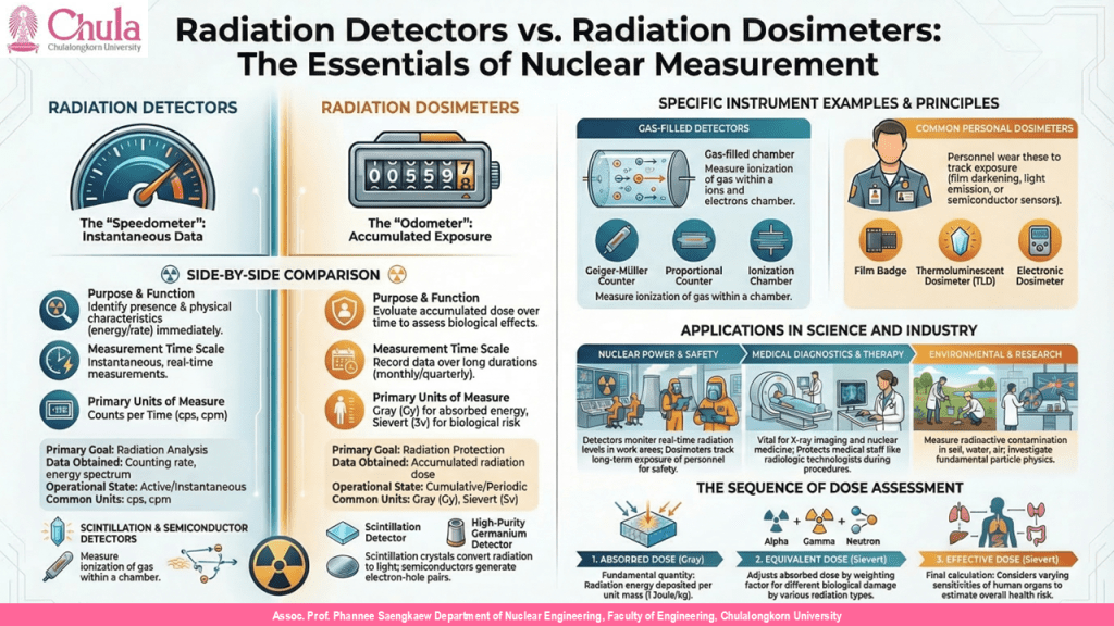
From a conceptual perspective, radiation measurement can generally be divided into two major approaches: radiation detection and radiation dosimetry. Radiation detection focuses on identifying the presence of radiation at a given moment and analyzing its physical characteristics, such as energy and counting rate. In contrast, radiation dosimetry focuses on evaluating the amount of radiation received by a person or object over a period of time, primarily for the purpose of assessing biological effects and ensuring radiation protection.
The instruments used for these two purposes serve clearly different roles. Radiation detectors are designed to detect and analyze radiation, whereas radiation dosimeters are used to measure accumulated radiation dose received by individuals for radiation protection purposes.
2 Conceptual Framework of Radiation Measurement Systems
Radiation measurement systems consist of more than a simple detection device. Instead, they are composed of multiple components that operate together in a sequence of processes, beginning with the physical interaction between radiation and matter and ending with the interpretation of measurement results.
In general, radiation measurement systems can be considered as having three main levels: the detection level, the instrumentation level, and the radiation assessment level.
At the first level, the radiation detector performs the task of detecting interactions between radiation and matter. When ionizing radiation enters a material, its energy is transferred to atoms or molecules within that material. This transfer of energy can produce several physical processes, including ionization, excitation of atomic energy levels, or the creation of electron–hole pairs in semiconductor materials.
At the second level, the initial signals produced by these interactions are processed through electronic systems that amplify, shape, and convert them into digital signals. Electronic processing plays an essential role in increasing signal strength and reducing background noise in order to obtain accurate measurement data.
At the final level, the resulting data are used for further analysis. Such analysis may include constructing radiation energy spectra, identifying radioactive isotopes, or calculating the radiation dose received by individuals.
3 Radiation Detectors
3.1 Definition and Role
A radiation detector is a device used to detect the presence of radiation by exploiting the interaction between radiation and matter to produce measurable signals. The detector acts as a transducer that converts radiation energy into signals that can be analyzed by measurement systems.
The role of radiation detectors extends beyond merely identifying the presence of radiation. They are also used to measure important characteristics of radiation, including energy, intensity, and type of radiation. Such information is essential for nuclear analysis, radioactive isotope identification, and the study of radiation–matter interactions.
3.2 Fundamental Principles of Radiation Detection
When ionizing radiation travels through matter, it transfers energy to atoms or molecules within the material. Several processes may occur during this interaction, including ionization of atoms, excitation of electrons, or the creation of electron–hole pairs in semiconductor materials.
These physical processes form the basis for radiation detection. For example, ionization occurring within gases can produce electrical currents, excitation within scintillation crystals can produce light, and electron–hole pairs generated within semiconductors can produce electrical pulses.
3.3 Types of Radiation Detectors
Radiation detectors can be classified according to the physical mechanism used for detection. Major categories include gas-filled detectors, scintillation detectors, and semiconductor detectors.
- Gas-filled detectors operate based on the ionization of gas within a detection chamber. Common examples include ionization chambers, proportional counters, and Geiger–Müller counters.
- Scintillation detectors utilize materials that convert radiation energy into light. The emitted light is then detected by light-sensitive devices such as photomultiplier tubes or photodiodes.
- Semiconductor detectors employ semiconductor materials such as silicon or germanium. When radiation interacts with these materials, electron–hole pairs are generated and collected as electrical signals.
4 Radiation Dosimeters
4.1 Definition
Radiation dosimeters are devices used to measure the accumulated radiation dose received by a person or object over a certain period of time. These instruments play a crucial role in radiation protection systems because they enable the evaluation of radiation exposure risks for workers and the general public.
In environments where radiation sources are present—such as nuclear power plants, hospitals, or nuclear laboratories—personnel are typically required to wear personal dosimeters. These devices allow monitoring of the radiation dose received during work activities.
The information recorded by dosimeters is analyzed periodically, often on a monthly or yearly basis, in order to determine whether radiation exposure remains within established safety limits.
4.2 Radiation Quantities Used in Dose Assessment
Assessing the effects of radiation on humans requires several radiation quantities, because different types of radiation produce different biological effects.
- The absorbed dose represents the amount of radiation energy absorbed per unit mass of material. The unit used for absorbed dose is the gray (Gy).
- The equivalent dose takes into account the type of radiation by applying radiation weighting factors. The unit used is the sievert (Sv).
- The effective dose further considers the varying sensitivities of different organs and tissues in the human body and is also expressed in sieverts.
5 Comparison between Radiation Detectors and Radiation Dosimeters
| Aspect | Radiation Detector | Radiation Dosimeter |
| Purpose | Detect radiation | Measure accumulated dose |
| Time scale | Instantaneous measurement | Accumulated over time |
| Information obtained | Counting rate, energy spectrum | Radiation dose |
| Units | Counts per time | Gray, Sievert |
| Application | Radiation analysis | Radiation protection |
6 Types of Detectors and Dosimeters
| Device Category | Example Instrument | Principle |
| Gas detectors | Ionization chamber | Gas ionization |
| Proportional counter | Charge multiplication | |
| Geiger–Müller counter | Avalanche discharge | |
| Scintillation detectors | Scintillation crystal | Conversion of radiation to light |
| Semiconductor detectors | Silicon detector | Electron–hole pair generation |
| High-purity germanium detector | High-resolution energy spectroscopy | |
| Radiation dosimeters | Film badge | Film darkening |
| Thermoluminescent dosimeter | Light emission upon heating | |
| Electronic dosimeter | Semiconductor sensor |
7 Roles of Radiation Detection Systems
Radiation detection systems play important roles in many scientific and technological fields, including radiation protection, nuclear medicine, environmental monitoring, and nuclear physics research.
In nuclear power plants, radiation monitoring systems are used to measure radiation levels in work areas and to track radiation exposure of personnel. In medicine, radiation detection systems are used for diagnostic imaging and radiation therapy. In environmental monitoring, radiation detection systems are used to measure radioactive contamination in soil, water, and air.
8 Relationship between Radiation Quantities for Human Dose Assessment
When evaluating the effects of radiation on humans, scientists consider not only the energy absorbed from radiation but also the type of radiation and the sensitivity of different organs in the human body. Therefore, several radiation quantities are defined in order to properly evaluate the biological effects of radiation exposure.
The relationship between these quantities can generally be expressed in the following sequence: Absorbed Dose → Equivalent Dose → Effective Dose
- Absorbed Dose
The absorbed dose is the most fundamental quantity used in radiation measurement. It represents the amount of radiation energy deposited per unit mass of material.
The unit of absorbed dose is the gray (Gy), defined as one joule of radiation energy absorbed per kilogram of matter.
Absorbed dose is widely used in radiation physics and radiation energy analysis. However, it does not fully represent the biological effects of radiation because different types of radiation cause different levels of biological damage.
- Equivalent Dose
Since different types of radiation produce different biological effects, the concept of equivalent dose was introduced. Equivalent dose is calculated by multiplying the absorbed dose by a radiation weighting factor.
The radiation weighting factor reflects the relative biological effectiveness of different types of radiation. For example, alpha particles typically produce greater biological damage than gamma radiation for the same absorbed dose.
The unit of equivalent dose is the sievert (Sv).
- Effective Dose
Even after considering the type of radiation, different organs and tissues in the human body exhibit different sensitivities to radiation. For instance, bone marrow, lungs, and reproductive organs are generally more sensitive to radiation exposure.
To account for these differences, the concept of effective dose was introduced. Effective dose incorporates both radiation weighting factors and tissue weighting factors to estimate the overall risk of radiation exposure to the human body.
Effective dose is also expressed in sieverts and forms the basis for radiation protection standards and dose limits.
9 Personal Radiation Monitoring Systems
In environments where radiation sources are present—such as nuclear power plants, hospitals, or nuclear laboratories—it is essential to monitor the radiation dose received by personnel. Such monitoring systems are known as personal radiation monitoring systems.
Workers who handle radiation sources typically wear personal dosimeters throughout their working hours. These devices are usually worn on the chest or waist in order to estimate the radiation dose received by the body.
The data recorded by personal dosimeters are periodically collected and analyzed, often monthly or quarterly. If the accumulated radiation dose approaches established limits, operational adjustments or protective measures may be implemented to ensure that exposure remains within safe levels i.e. < 20 mSv/y for radiation workers and < 1 mSv/y for general public.
In addition to monitoring individual exposure, dosimeters may also be placed in specific locations within facilities—such as reactor buildings or laboratories—to study the spatial distribution of radiation and evaluate long-term radiation accumulation in the working environment.
10 Radiation Dose Limits
The establishment of radiation dose limits is an essential component of radiation protection systems. The fundamental principle is to limit the amount of radiation that individuals may receive to levels that do not pose unacceptable health risks.
Radiation dose limits are established by international organizations such as the International Commission on Radiological Protection (ICRP). Different limits are specified for occupational radiation workers and for members of the general public.
Workers who are professionally exposed to radiation may receive higher doses than the general public because their exposure is carefully controlled and monitored. Nevertheless, exposure must remain within the established limits, and appropriate radiation protection measures must be implemented.
A key concept in radiation protection is the principle that radiation exposure should be kept as low as reasonably achievable (ALARA), taking into account technological, economic, and social factors. This principle helps minimize long-term health risks associated with radiation exposure.
11 Practical Applications of Radiation Dosimeters
Radiation dosimeters are used in a wide range of practical applications.
- In nuclear power plants, dosimeters are used to monitor the radiation doses received by workers operating in controlled areas. These measurements help ensure that maintenance operations and routine tasks are carried out safely in radiation environments.
- In medical settings, radiation dosimeters are used to monitor the radiation exposure of medical personnel such as radiologic technologists and physicians who work with diagnostic imaging equipment or radiation therapy systems.
- In nuclear research laboratories, dosimeters help track radiation distribution within experimental environments and support the implementation of radiation safety protocols.
12 Overall Perspective of Radiation Detection Systems
From the discussions presented in this chapter, it is evident that radiation measurement systems consist of two principal categories of instruments: radiation detectors and radiation dosimeters.
- Radiation detectors are used to detect and analyze radiation at a given moment, such as measuring radiation counting rates or analyzing radiation energy spectra.
- Radiation dosimeters are used to monitor accumulated radiation exposure received by individuals or objects over time.
Both types of systems play critical roles in radiation protection, nuclear research, and the application of radiation in fields such as medicine, industry, and environmental monitoring.
A thorough understanding of the principles and functions of these instruments is therefore essential for scientists and nuclear engineers who design and operate radiation measurement systems in a safe and effective manner.
*************************************************************
การตรวจวัดรังสีและการวัดปริมาณรังสีที่ได้รับ
(Radiation Detection and Radiation Dosimetry)
1 บทนำ
การตรวจวัดรังสีเป็นองค์ประกอบพื้นฐานของวิทยาศาสตร์และวิศวกรรมนิวเคลียร์ เนื่องจากรังสีก่อให้เกิดไอออนเป็นปรากฏการณ์ทางกายภาพที่ไม่สามารถรับรู้ได้โดยตรงด้วยประสาทสัมผัสของมนุษย์ รังสีไม่มีสี ไม่มีกลิ่น และไม่สามารถมองเห็นได้ด้วยตาเปล่า ดังนั้น การศึกษาการมีอยู่ของรังสี การวัดปริมาณรังสี และการวิเคราะห์พลังงานของรังสีจึงต้องอาศัยเครื่องมือเฉพาะทางที่สามารถตรวจวัดผลของการทำอันตรกิริยาระหว่างรังสีกับสสาร และแปลงพลังงานของรังสีให้เป็นสัญญาณที่สามารถวัด วิเคราะห์ และตีความได้
ในทางปฏิบัติ การตรวจวัดรังสีมีบทบาทสำคัญในหลายสาขาของวิทยาศาสตร์และเทคโนโลยี ตัวอย่างเช่น ในโรงไฟฟ้านิวเคลียร์ ระบบตรวจวัดรังสีถูกใช้เพื่อติดตามระดับรังสีในพื้นที่ทำงานและควบคุมความปลอดภัยของการดำเนินงาน ในทางการแพทย์ ระบบตรวจวัดรังสีถูกนำไปใช้ในการวินิจฉัยและการรักษาโรค เช่น การถ่ายภาพทางการแพทย์ด้วยรังสีเอกซ์ การถ่ายภาพด้วยเวชศาสตร์นิวเคลียร์ และการรักษาด้วยรังสีรักษา นอกจากนี้การตรวจวัดรังสียังมีบทบาทสำคัญในการเฝ้าระวังด้านสิ่งแวดล้อม การวิเคราะห์ตัวอย่างทางธรณีวิทยา และการศึกษาปฏิกิริยามูลฐานในฟิสิกส์นิวเคลียร์และฟิสิกส์อนุภาค
เมื่อพิจารณาในเชิงแนวคิด การวัดรังสีสามารถแบ่งออกเป็นสองแนวทางหลัก ได้แก่ การตรวจวัดรังสี และ การวัดปริมาณรังสีที่ได้รับ การตรวจวัดรังสีมุ่งเน้นการตรวจพบการมีอยู่ของรังสีในช่วงเวลาหนึ่ง รวมทั้งการศึกษาคุณสมบัติทางกายภาพของรังสี เช่น พลังงานและอัตราการเกิดรังสี ในทางตรงกันข้าม การวัดปริมาณรังสีที่ได้รับมุ่งเน้นการประเมินปริมาณรังสีที่บุคคลหรือวัตถุได้รับในช่วงเวลาหนึ่ง เพื่อใช้ในการประเมินผลเสียทางชีวภาพของรังสีต่อสิ่งมีชีวิต
เครื่องมือที่ใช้ในสองกรณีนี้มีบทบาทแตกต่างกันอย่างชัดเจน ได้แก่ หัววัดรังสี (Radiation detector) ซึ่งใช้ตรวจวัดและวิเคราะห์รังสี และ อุปกรณ์วัดปริมาณรังสี (Radiation dosimeter) ซึ่งใช้ประเมินปริมาณรังสีที่บุคคลได้รับเพื่อวัตถุประสงค์ด้านความปลอดภัยทางรังสี
2 กรอบแนวคิดของระบบการวัดรังสี
ระบบการวัดรังสีไม่ได้ประกอบด้วยเพียงอุปกรณ์ตรวจวัดเพียงอย่างเดียว แต่เป็นระบบที่ประกอบด้วยองค์ประกอบหลายส่วนที่ทำงานร่วมกันเป็นลำดับขั้น ตั้งแต่กระบวนการตรวจวัดทางกายภาพไปจนถึงการวิเคราะห์ข้อมูล
โดยทั่วไปสามารถแบ่งระบบการวัดรังสีออกเป็นสามระดับสำคัญ ได้แก่ ระดับการตรวจวัด ระดับระบบเครื่องมือ และระดับการประเมินผลทางรังสี
- ในระดับแรก หัววัดรังสีทำหน้าที่ตรวจวัดการทำอันตรกิริยาระหว่างรังสีกับสสาร เมื่อรังสีก่อให้เกิดไอออนผ่านเข้าสู่สสาร พลังงานของรังสีจะถูกถ่ายโอนไปยังอะตอมหรือโมเลกุลของวัสดุ ทำให้เกิดกระบวนการต่าง ๆ เช่น การแตกตัวเป็นไอออน การกระตุ้นสถานะพลังงานของอะตอม หรือการสร้างคู่ประจุในวัสดุกึ่งตัวนำ
- ในระดับที่สอง สัญญาณเริ่มต้นที่เกิดขึ้นจากการทำอันตรกิริยาดังกล่าวจะถูกส่งผ่านระบบอิเล็กทรอนิกส์เพื่อขยาย ปรับแต่ง และแปลงเป็นสัญญาณดิจิทัล ระบบอิเล็กทรอนิกส์มีบทบาทสำคัญในการเพิ่มขนาดของสัญญาณและลดสัญญาณรบกวน
- ในระดับสุดท้าย ข้อมูลจากระบบตรวจวัดจะถูกนำไปใช้ในการวิเคราะห์ เช่น การสร้างสเปกตรัมพลังงานของรังสี การระบุชนิดของไอโซโทป หรือการคำนวณปริมาณรังสีที่บุคคลได้รับ
3 หัววัดรังสี
3.1 ความหมายและบทบาท
หัววัดรังสีเป็นอุปกรณ์ที่ใช้ตรวจวัดการมีอยู่ของรังสี โดยอาศัยการทำอันตรกิริยาระหว่างรังสีกับสสารตัวกลางที่เคลื่อนที่ผ่าน เพื่อสร้างสัญญาณที่สามารถตรวจวัดได้ หัววัดรังสีทำหน้าที่เป็นตัวแปลงพลังงานจากพลังงานของรังสีให้กลายเป็นสัญญาณที่สามารถนำไปวิเคราะห์ได้ บทบาทของหัววัดรังสีไม่ได้จำกัดอยู่เพียงการตรวจพบการมีอยู่ของรังสีเท่านั้น แต่ยังรวมถึงการวัดวิเคราะห์คุณสมบัติเฉพาะของรังสี เช่น พลังงาน อัตราการเกิดรังสี และชนิดของรังสี ข้อมูลเหล่านี้มีความสำคัญต่อการวิเคราะห์ทางนิวเคลียร์ การระบุไอโซโทปกัมมันตรังสี และการศึกษาการทำอันตรกิริยาของรังสีกับสสาร
3.2 หลักการพื้นฐานของการตรวจวัดรังสี
เมื่อรังสีก่อให้เกิดไอออนเคลื่อนที่ผ่านสสาร จะเกิดการถ่ายเทพลังงานให้กับอะตอมหรือโมเลกุลของวัสดุ กระบวนการดังกล่าวสามารถเกิดขึ้นได้หลายรูปแบบ เช่น การแตกตัวเป็นไอออนของอะตอม การกระตุ้นสถานะพลังงานของอิเล็กตรอน หรือการสร้างคู่อิเล็กตรอนและโฮลในวัสดุกึ่งตัวนำ
ผลของการทำอันตรกิริยาเหล่านี้สามารถนำมาใช้ในการตรวจวัดรังสีได้ ตัวอย่างเช่น การแตกตัวเป็นไอออนของก๊าซสามารถสร้างกระแสไฟฟ้า การกระตุ้นผลึกซินทิลเลเตอร์สามารถทำให้เกิดการปล่อยแสง และการสร้างคู่ประจุในวัสดุกึ่งตัวนำสามารถสร้างพัลส์สัญญาณไฟฟ้าได้
3.3 ประเภทของหัววัดรังสี
หัววัดรังสีสามารถแบ่งออกเป็นหลายประเภทตามกลไกการตรวจวัด คือ
- หัววัดรังสีชนิดก๊าซอาศัยการแตกตัวเป็นไอออนของก๊าซภายในหลอดตรวจวัด ตัวอย่างที่ใช้กันอย่างแพร่หลาย ได้แก่ ห้องไอออไนเซชัน หลอดสัดส่วน และหลอดไกเกอร์–มึลเลอร์
- หัววัดรังสีชนิดซินทิลเลเตอร์ใช้ผลึกที่สามารถเปลี่ยนพลังงานของรังสีให้เป็นแสง จากนั้นแสงจะถูกตรวจวัดด้วยอุปกรณ์ตรวจแสง
- หัววัดรังสีชนิดสารกึ่งตัวนำใช้วัสดุ เช่น ซิลิคอนหรือเจอร์เมเนียม ซึ่งสามารถสร้างคู่อิเล็กตรอนและโฮลเมื่อได้รับพลังงานจากรังสี
4 อุปกรณ์วัดปริมาณรังสี
4.1 ความหมาย
อุปกรณ์วัดปริมาณรังสีเป็นอุปกรณ์ที่ใช้สำหรับวัดปริมาณรังสีที่บุคคลหรือวัตถุได้รับสะสมในช่วงเวลาหนึ่ง อุปกรณ์ชนิดนี้มีบทบาทสำคัญในระบบการป้องกันอันตรายจากรังสี เนื่องจากสามารถใช้ประเมินความเสี่ยงจากการได้รับรังสีของบุคลากรหรือประชาชน
ในงานที่เกี่ยวข้องกับการใช้รังสี เช่น โรงไฟฟ้านิวเคลียร์ โรงพยาบาล หรือห้องปฏิบัติการนิวเคลียร์ บุคลากรมักต้องสวมอุปกรณ์วัดปริมาณรังสีติดตัวไว้ เพื่อให้สามารถติดตามปริมาณรังสีที่ได้รับระหว่างการทำงาน ข้อมูลจากอุปกรณ์วัดปริมาณรังสีจะถูกนำไปวิเคราะห์เป็นระยะ เช่น รายเดือนหรือรายปี เพื่อประเมินว่าการได้รับรังสีอยู่ภายในขอบเขตที่ปลอดภัยหรือไม่
4.2 ปริมาณรังสีที่ใช้ในการประเมิน
การประเมินผลเสียของรังสีต่อมนุษย์ต้องอาศัยปริมาณรังสีหลายชนิด เนื่องจากรังสีแต่ละชนิดมีผลเสียต่อเนื้อเยื่อแตกต่างกัน
- ปริมาณรังสีดูดกลืน ใช้วัดพลังงานของรังสีที่ถูกดูดกลืนต่อหน่วยมวลของสสาร หน่วยที่ใช้คือ เกรย์
- ปริมาณรังสีสมมูล จากที่คำนึงถึงความแตกต่างของชนิดของรังสี โดยใช้ปัจจัยถ่วงน้ำหนักของรังสี หน่วยที่ใช้คือ ซีเวิร์ต
- ปริมาณรังสียังผล ที่ส่งผลเสียต่อร่างกายจากที่คำนึงถึงความไวต่อรังสีของอวัยวะต่าง ๆ ของร่างกาย โดยใช้ปัจจัยถ่วงน้ำหนักของอวัยวะแต่ละประเภทนั้น และมีหน่วยเป็น ซีเวิร์ต
5 ตารางเปรียบเทียบหัววัดรังสีและอุปกรณ์วัดปริมาณรังสี
| ประเด็น | หัววัดรังสี | อุปกรณ์วัดปริมาณรังสี |
| วัตถุประสงค์ | ตรวจวัดรังสี | วัดปริมาณรังสีที่ได้รับ |
| ช่วงเวลา | แบบทันที | สะสมตามเวลา |
| ข้อมูลที่ได้ | อัตราการนับ พลังงาน | ปริมาณรังสี |
| หน่วยวัด | การนับต่อเวลา | เกรย์ ซีเวิร์ต |
| การใช้งาน | เครื่องมือวิเคราะห์รังสี | การป้องกันรังสี |
6 ตารางชนิดของเครื่องตรวจวัดและอุปกรณ์วัดปริมาณรังสี
| หมวดอุปกรณ์ | ตัวอย่างเครื่องมือ | หลักการ |
| หัววัดรังสีชนิดก๊าซ | ห้องไอออไนเซชัน | การแตกตัวเป็นไอออน |
| หลอดสัดส่วน | การขยายประจุ | |
| หลอดไกเกอร์–มึลเลอร์ | การปลดปล่อยประจุ | |
| หัววัดซินทิลเลเตอร์ | ผลึกซินทิลเลเตอร์ | การเปลี่ยนพลังงานเป็นแสง |
| หัววัดกึ่งตัวนำ | ซิลิคอน | การสร้างคู่อิเล็กตรอนโฮล |
| เจอร์เมเนียมบริสุทธิ์สูง | การวิเคราะห์สเปกตรัมพลังงาน | |
| อุปกรณ์วัดปริมาณรังสี | ฟิล์มโดซิมิเตอร์ | การเปลี่ยนสีของฟิล์ม |
| โดซิมิเตอร์เทอร์โมลูมิเนสเซนซ์ | การปล่อยแสงเมื่อให้ความร้อน | |
| โดซิมิเตอร์อิเล็กทรอนิกส์ | เซนเซอร์กึ่งตัวนำ |
7 บทบาทของระบบตรวจวัดรังสี
ระบบตรวจวัดรังสีมีบทบาทสำคัญในหลายสาขาของวิทยาศาสตร์และเทคโนโลยี เช่น การป้องกันรังสี เวชศาสตร์นิวเคลียร์ การเฝ้าระวังสิ่งแวดล้อม และการวิจัยทางฟิสิกส์นิวเคลียร์
ในโรงไฟฟ้านิวเคลียร์ ระบบตรวจวัดรังสีใช้เพื่อติดตามระดับรังสีในพื้นที่ทำงานและติดตามการได้รับปริมาณรังสีของบุคลากร ในทางการแพทย์ ระบบตรวจวัดรังสีถูกใช้ในการสร้างภาพทางการแพทย์และการรักษาด้วยรังสี ส่วนในงานสิ่งแวดล้อม ระบบตรวจวัดรังสีถูกใช้ติดตามระดับกัมมันตภาพรังสีในดิน น้ำ และอากาศ
8 ความสัมพันธ์ของปริมาณรังสีที่ใช้ในการประเมินผลเสียต่อมนุษย์
ในการประเมินผลเสียของรังสีต่อมนุษย์ นักวิทยาศาสตร์ไม่ได้พิจารณาเพียงพลังงานของรังสีที่ถูกดูดกลืนเท่านั้น แต่ยังต้องคำนึงถึงชนิดของรังสีและความไวของอวัยวะต่าง ๆ ในร่างกายด้วย ดังนั้นจึงมีการกำหนดปริมาณรังสีหลายชนิดเพื่อใช้ในการประเมินผลเสียทางชีวภาพของรังสีอย่างเหมาะสม
โดยทั่วไป ความสัมพันธ์ของปริมาณรังสีสามารถอธิบายได้เป็นลำดับขั้นดังนี้
ปริมาณรังสีดูดกลืน → ปริมาณรังสีสมมูล → ปริมาณรังสียังผล
ปริมาณรังสีดูดกลืน
ปริมาณรังสีดูดกลืนเป็นค่าพื้นฐานที่สุดในการวัดรังสี โดยแสดงถึงพลังงานของรังสีที่ถูกดูดกลืนต่อหน่วยมวลของสสาร ปริมาณนี้สะท้อนการถ่ายเทพลังงานจากรังสีเข้าสู่เนื้อเยื่อหรือวัสดุ หน่วยของปริมาณรังสีดูดกลืนคือ เกรย์ (Gray) ซึ่งหมายถึงพลังงานหนึ่งจูลที่ถูกดูดกลืนต่อมวลหนึ่งกิโลกรัมของสสาร ปริมาณนี้มีความสำคัญในงานฟิสิกส์รังสีและการวิเคราะห์พลังงานของรังสี อย่างไรก็ตาม ค่านี้ยังไม่สามารถสะท้อนผลเสียทางชีวภาพของรังสีได้อย่างครบถ้วน เนื่องจากรังสีแต่ละชนิดมีความสามารถในการทำลายเนื้อเยื่อแตกต่างกัน
ปริมาณรังสีสมมูล
เพื่อคำนึงถึงความแตกต่างของผลเสียจากรังสีแต่ละชนิด จึงมีการกำหนดปริมาณรังสีสมมูล ซึ่งคำนวณจากปริมาณรังสีดูดกลืนคูณด้วยปัจจัยถ่วงน้ำหนักของชนิดรังสี ปัจจัยนี้สะท้อนความสามารถของรังสีแต่ละชนิดในการทำลายเนื้อเยื่อ ตัวอย่างเช่น อนุภาคแอลฟามีความสามารถในการก่อให้เกิดความเสียหายต่อเนื้อเยื่อสูงกว่ารังสีแกมมา หน่วยของปริมาณรังสีสมมูลคือ ซีเวิร์ต (Sievert)
ปริมาณรังสียังผล
แม้ว่าปริมาณรังสีสมมูลจะคำนึงถึงชนิดของรังสีแล้ว แต่ร่างกายมนุษย์ประกอบด้วยอวัยวะหลายชนิดซึ่งมีความไวต่อรังสีแตกต่างกัน เช่น ไขกระดูก ปอด หรือระบบอวัยวะสืบพันธุ์ ดังนั้นจึงมีการกำหนดปริมาณรังสีที่คำนึงถึงความไวของอวัยวะต่าง ๆ ในร่างกาย เพื่อใช้ในการประเมินความเสี่ยงโดยรวมต่อสุขภาพของมนุษย์ ปริมาณนี้เรียกว่า ปริมาณรังสียังผล ปริมาณดังกล่าวใช้หน่วยเป็นซีเวิร์ตเช่นเดียวกับปริมาณรังสีสมมูล และถูกนำไปใช้เป็นพื้นฐานในการกำหนดมาตรฐานความปลอดภัยทางรังสี
9 ระบบติดตามปริมาณรังสีของบุคลากร
ในสถานที่ที่มีการใช้รังสี เช่น โรงไฟฟ้านิวเคลียร์ โรงพยาบาล หรือห้องปฏิบัติการนิวเคลียร์ จำเป็นต้องมีระบบติดตามปริมาณรังสีที่บุคลากรได้รับ ระบบดังกล่าวเรียกว่า ระบบติดตามปริมาณรังสีบุคคล
บุคลากรที่ทำงานกับแหล่งกำเนิดรังสีมักต้องสวมอุปกรณ์วัดปริมาณรังสีติดตัวอยู่ตลอดระยะเวลาการทำงาน อุปกรณ์นี้มักติดตั้งไว้บริเวณหน้าอกหรือบริเวณเอว เพื่อให้สามารถประเมินปริมาณรังสีที่ร่างกายได้รับได้อย่างเหมาะสม
ข้อมูลจากอุปกรณ์วัดปริมาณรังสีจะถูกเก็บรวบรวมและวิเคราะห์เป็นระยะ เช่น รายเดือนหรือรายไตรมาส หากพบว่าปริมาณรังสีที่บุคลากรได้รับเข้าใกล้ขีดจำกัดที่กำหนด ก็จะมีการปรับเปลี่ยนลักษณะการทำงานหรือมาตรการป้องกันเพื่อให้มั่นใจว่าการได้รับรังสีอยู่ในระดับที่ปลอดภัย
นอกจากการติดตามปริมาณรังสีของบุคลากรแล้ว ในบางกรณียังมีการใช้อุปกรณ์วัดปริมาณรังสีเพื่อศึกษาการกระจายของรังสีในพื้นที่ เช่น การติดตั้งอุปกรณ์ในจุดต่าง ๆ ของอาคารปฏิกรณ์หรือสถานที่ปฏิบัติงาน เพื่อประเมินการสะสมของรังสีในระยะยาว
10 แนวคิดขีดจำกัดการได้รับรังสี
การกำหนดขีดจำกัดการได้รับรังสี (Radiation Dose Limitation) เป็นองค์ประกอบสำคัญของระบบการป้องกันอันตรายจากรังสี หลักการสำคัญคือการจำกัดปริมาณรังสีที่บุคคลสามารถได้รับโดยไม่ก่อให้เกิดความเสี่ยงต่อสุขภาพในระดับที่ยอมรับไม่ได้
ขีดจำกัดการได้รับรังสีถูกกำหนดโดยองค์กรระหว่างประเทศ เช่น คณะกรรมาธิการระหว่างประเทศว่าด้วยการป้องกันรังสี (International Commission on Radiological Protection) โดยมีการกำหนดค่าขีดจำกัดที่แตกต่างกันสำหรับบุคลากรที่ทำงานกับรังสีและประชาชนทั่วไป
บุคลากรที่ทำงานกับรังสีสามารถได้รับรังสีในระดับที่สูงกว่าประชาชนทั่วไปได้ เนื่องจากมีการควบคุมและติดตามการได้รับรังสีอย่างใกล้ชิด อย่างไรก็ตาม การได้รับรังสีต้องอยู่ภายในขอบเขตที่กำหนด และต้องมีการใช้มาตรการป้องกันรังสีอย่างเหมาะสม กล่าวอย่างง่ายคือโดยเฉลี่ยไม่เกิน 20 mSv ต่อปี และประชาชยทั่วไปไม่เกิน 1 mSv ต่อปี
แนวคิดสำคัญในการป้องกันรังสีคือการทำให้การได้รับรังสี ต่ำที่สุดเท่าที่สามารถทำได้อย่างสมเหตุสมผล โดยคำนึงถึงปัจจัยทางเทคนิค เศรษฐกิจ และสังคม หลักการนี้ช่วยลดความเสี่ยงจากการได้รับรังสีในระยะยาว
11 การประยุกต์ใช้อุปกรณ์วัดปริมาณรังสีในงานจริง
อุปกรณ์วัดปริมาณรังสีถูกนำไปใช้ในหลากหลายสาขา
- ในโรงไฟฟ้านิวเคลียร์ อุปกรณ์เหล่านี้ใช้เพื่อติดตามปริมาณรังสีที่บุคลากรได้รับระหว่างการทำงานในพื้นที่ควบคุม ข้อมูลดังกล่าวช่วยให้สามารถจัดการงานบำรุงรักษาและการปฏิบัติงานในพื้นที่ที่มีรังสีได้อย่างปลอดภัย
- ในทางการแพทย์ อุปกรณ์วัดปริมาณรังสีใช้เพื่อติดตามการได้รับรังสีของบุคลากรทางการแพทย์ เช่น นักรังสีเทคนิคหรือแพทย์ที่ทำงานกับเครื่องเอกซเรย์หรือรังสีรักษา
- ในงานวิจัยทางนิวเคลียร์ อุปกรณ์วัดปริมาณรังสีช่วยให้สามารถติดตามการกระจายของรังสีในห้องทดลองและประเมินความปลอดภัยของการทดลอง
12 สรุปภาพรวมของระบบตรวจวัดรังสี
จากเนื้อหาทั้งหมดจะเห็นได้ว่าระบบตรวจวัดรังสีประกอบด้วยเครื่องมือสองกลุ่มหลัก ได้แก่ หัววัดรังสี และ อุปกรณ์วัดปริมาณรังสี
หัววัดรังสีทำหน้าที่ตรวจวัดและวิเคราะห์รังสีในช่วงเวลาหนึ่ง เช่น การวัดอัตราการนับของรังสีหรือการวิเคราะห์สเปกตรัมพลังงานของรังสี ส่วนอุปกรณ์วัดปริมาณรังสีใช้ในการติดตามปริมาณรังสีที่บุคคลหรือวัตถุได้รับในช่วงเวลาหนึ่ง
ทั้งสองระบบมีบทบาทสำคัญต่อความปลอดภัยทางรังสี การวิจัยทางนิวเคลียร์ และการประยุกต์ใช้รังสีในด้านต่าง ๆ เช่น การแพทย์ อุตสาหกรรม และการเฝ้าระวังผลผระทบต่อสิ่งแวดล้อม
ความเข้าใจในหลักการทำงานของเครื่องมือหรืออุปกรณ์วัดเหล่านี้จึงเป็นพื้นฐานสำคัญสำหรับนักวิทยาศาสตร์และวิศวกรนิวเคลียร์ในการออกแบบและใช้งานระบบตรวจวัดรังสีอย่างถูกต้องและมีประสิทธิภาพ

ใส่ความเห็น